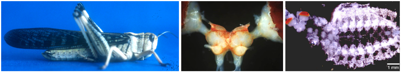
We are interested in structural and functional neuronal plasticity of the insect nervous system. The nervous system is able to respond and adapt to a large variety of external factors during development and adult life, like environmental changes, injury, infection, or toxins, in order to ensure survival and reproduction of the animal. We are interested in the role of neuromodulatory transmitters in these adaptation processes, with a focus on biogenic amines (serotonin and octopamine), and the unusual gaseous transmitter nitric oxide (NO). In the following we will give some examples of our projects.
Literature
Stern M (1999) Octopamine in the locust brain: Cellular distribution and functional significance in an arousal mechanism. Micr. Res. Tech. 45:135-141.
Stern M, Bicker G (2010) NO as a regulator of neuronal motility and regeneration in the locust nervous system. J. Insect Physiol. 56:958-965.
Bicker G, Stern M (2020) Structural and functional plasticity in the regenerating olfactory system of the migratory locust. Front. Physiol. 608661. doi: 10.3389/fphys.2020.608661
We employ various histological and biochemical techniques to localize and visualize and to interfere with this highly reactive and rather elusive molecule and its signaling pathways. Our standard model organism is the locust (Locusta migratoria) which is very useful because of its large size and robustness, but we also work with Drosophila, mosquitos, other invertebrates, and vertebrate cell lines. We could show that in the locust CNS, NO is produced in response to nerve injury and that it facilitates nerve regeneration.
Literature
Stern M, Bicker G (2008) Nitric oxide regulates axonal regeneration in an insect embryonic CNS. Dev. Neurobiol. 68:295-308.
Stern M, Böger N, Eickhoff R, Kerßen U, Lorbeer C, Ziegler M, Martinelli GP, Holstein GR, Bicker G (2010) Development of nitrergic neurons in the nervous system of the locust embryo. J. Comp. Neurol. 518:1157–1175.
Scheiblich H, Roloff F, Singh V, Stangel M, Stern M, Bicker G (2014) Nitric oxide / cyclic GMP signaling regulates motility of a microglial cell line in vitro. Brain Res. 1564:9-21.
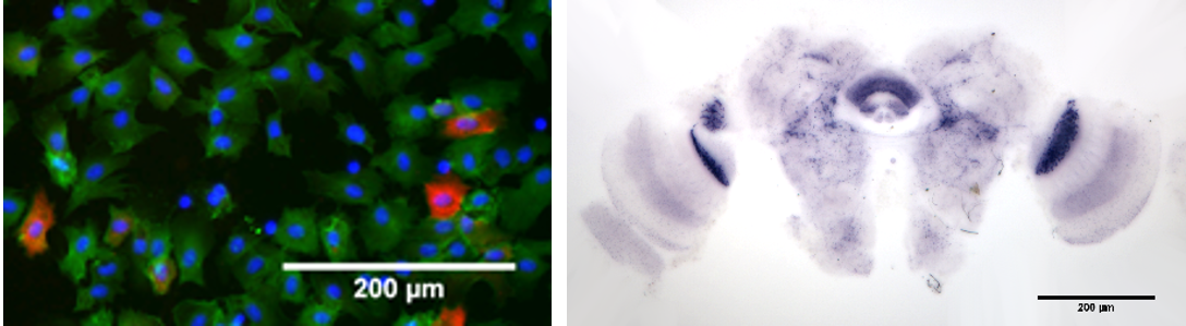
In a current DFG-funded project, we investigate the involvement of NO in neuro-immune interactions in collaboration with the group of Prof. Stephanie Becker, Institute of Parasitology. The objective is to elucidate possible implications of an infection (viral or bacterial) on the nervous system and behavior of insects, in particular the biting behavior of mosquitos carrying a pathogenic arbovirus. To this end, we measure NO-production in hemocytes, the insect’s immune cells, in response to pathogens, and quantify the behavior of infected vs. non-infected insects in a semi-automatic fashion.
Literature
Bergmann S, Gerhards JP, Schmitz A, Becker SC, Stern M (2021) NO synthesis in immune-challenged locust hemocytes and potential signaling to the CNS. Insects 12(10) 951. https://doi.org/10.3390/insects12100951
Bergmann S, Bohn MC, Dornbusch S, Becker SC, Stern M (2023) Influence of RVFV infection on olfactory perception and behavior in Drosophila melanogaster. Pathogens 12: 558. https://doi.org/10.3390/pathogens12040558
Bergmann S, Graf E, Hoffmann P, Becker SC, Stern M (2024) Localization of nitric oxide producing hemocytes in Aedes and Culex mosquitoes infected with bacteria. Cell Tissue Res (2024). https://doi.org/10.1007/s00441-024-03862-1
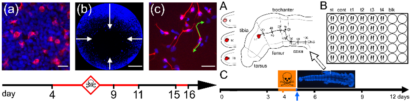
A further field of our research interests is developmental neurotoxicity (DNT). Exposure to pesticides or other toxins during in utero and early postnatal development can cause a wide range of neurological defects. In vivo testing of chemicals in vertebrates is cost-intensive. In collaboration with the virtual center for replacement of animal experiments, we are involved in the development of alternative methods to test the neurotoxic potential of chemicals. We employ a human neuronal precursor cell line (Ntera2) to develop standardized tests for toxin effects on fundamental processes of brain development like cell migration, neuronal differentiation, and neurite outgrowth. More advanced developmental processes, like axonal growth cone navigation along multiple morphogen gradients can hardly be addressed in simple cell culture systems. Making use of our expertise on the insect nervous system, we have recently developed a test method for DNT on axon navigation using embryonic locust limb bud pioneer neurons as a test system.
Literature
Stern M, Gierse A, Tan S, Bicker G (2014) Human Ntera-2 cells as a predictive in vitro test system for developmental neurotoxicity. Arch. Toxicol. 88:127–136.
Bergmann GA, Frömbling S, Joseph N, Bode K, Bicker G, Stern M (2019) An intact insect embryo for developmental neurotoxicity testing of directed axonal elongation. ALTEX 36(4):643-649. doi:10.14573/altex.1901292
Bode K, Bohn M, Reitmeier J, Betker P, Stern M, Bicker G (2020) A locust embryo as predictive developmental neurotoxicity testing system for pioneer axon pathway formation. Arch Toxicol. 94: 4099-4113. doi: 10.1007/s00204-020-02929-6.
Schmitz A, Dempewolf S, Tan S, Bicker G, Stern M (2021) Developmental neurotoxicity of fipronil and rotenone on a human neuronal in vitro test system. Neurotox. Res. https://doi.org/10.1007/s12640-021-00364-8
Stern M, Botha N, Cloete KJ, Maaza M, Tan S, Bicker G (2024) Neurotoxicity and developmental neurotoxicity of copper sulfide nanoparticles on a human neuronal in vitro test system. Int J Mol Sci 25 (11): 5650. https://doi.org/10.3390/ijms25115650
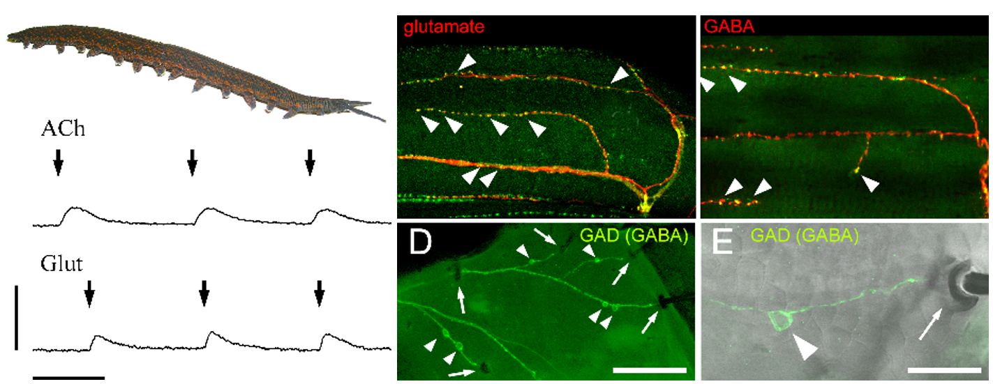
In a long-lasting project, we investigate the chemical neuroarchitecture of invertebrates as potential phylogenetic traits. In the last two decades, the use of molecular biological cues has led to major restructuring of phylogenetic trees, sometimes contradicting classical views based on morphology. Synaptic neurotransmitter equipment could provide additional independent information, here. Whereas vertebrates use acetylcholine (ACh) as the excitatory neuromuscular transmitter, insects and crustaceans use glutamate instead, with ACh as the transmitter of sensory neurons. In addition, several arthropods have GABAergic inhibitory motor neurons. We analyze a broad variety of arthropod and non-arthropod taxa in order to gain information about their motor and also sensory transmitters. We could show that the phylum Onychophora, at the base of the arthropod clade, uses both ACh and Glutamate as neuromuscular transmitters, and that centipedes have GABAergic sensory neurons.
Literature
Stern M, Bicker G (2008) Mixed cholinergic/glutamatergic neuromuscular innervation of Onychophora: A combined histochemical/electrophysiological study. Cell Tissue Res. 333:333-338.
Langeloh H, Wasser H, Richter N, Bicker G, Stern M (2018) Neuromuscular transmitter candidates of a centipede (Lithobius forficatus, Chilopoda). Front. Zool. 15:28
In all projects, we offer Bachelor theses, Master 3. semester research modules and Master theses.
If you are interested, contact Michael.Stern@tiho-hannover.de