One pathogen - many animals
How susceptible are pets and livestock to SARS-CoV-2? In a collaborative project, a team of researchers at the Institute of Pathology is testing whether and how the virus infects pets.
The virus pandemic triggered by SARS-CoV-2 is a zoonosis: Severe Acute Respiratory Syndrome Coronavirus 2 (SARS-CoV-2) originates from animals. It is unclear which and how many species it can infect so far. Especially where animals are kept in close contact with humans, it is important to know whether these species are susceptible to such a viral infection. However, little is known to date about whether pets can become infected with SARS-CoV-2.
A team of the Institute of Pathology of the TiHo is now looking into this in the project Domestic Animals as Potential Vectors for SARS-CoV-2 Transmission (ANI-CoV) together with colleagues from the Department of Infection Biology of the Leibniz Institute for Primate Research in Göttingen. In the project, the researchers are specifically looking at what happens at the cellular level in the respiratory tract of animals when they get into contact with the COVID-19 trigger. The trick is that animal experiments are not required. And it could also start right away, because at the Institute of Pathology, the team led by Professor Dr. Wolfgang Baumgärtner, PhD, had already acquired expertise and material to set up a tissue and cell bank with the financial support of the Lower Saxony Ministry of Science and Culture. The collection is part of the R2N - Replace and Reduce project from Lower Saxony, which focuses on animal-free basic biomedical research on respiratory infections in humans and animals. Now the researchers can use the tissue and cell cultures for their SARS-CoV-2 research. "So we already have material from common pet and farm animal species such as dogs, cats, pigs, ferrets, etc. in stock," explains research assistant Sandra Runft. Tissue cultures have been established from a total of eleven animal species so far. Since little is known about the different tissue structures in the respiratory tracts of the animal species, the careful characterization of the tissues is one of the tasks of the research team.
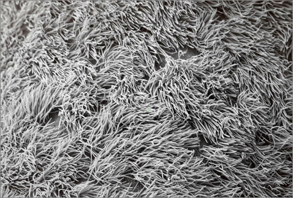
In ANI-CoV, the researchers are testing whether the wafer-thin tissue cultures can be infected with SARS-CoV-2. Not all cells are the same: "We want to study both the upper and lower respiratory tract," Runft explains, and enumerates: "Nasal mucosa, trachea, tracheal epithelial cells, the ciliated epithelial cells, lung tissue - from the nose to the lungs, there are different tissue types." They use a variety of ex vivo and in vitro culture systems, such as nasal mucosal explants, air-liquid interface cultures or precision-cut lung slices (PCLS).
Using the cell cultures, the next step is to identify the domestic species that are actually susceptible to SARS-CoV-2. The next step is to identify the cell types that react to the virus. The question is which tissue types in which animal species can actually be attacked by the virus and what modifications occur as a result. Whether cells die or tissue structures change can be tracked, for example, using a transmission electron microscope. "We also want to investigate whether we see differences between the cells of the different animal species," Runft explains, "and whether we can observe a reaction in the different immune cells." This is where the colleagues from Göttingen come in: Nadine Krüger, PhD, and her team are analyzing, among other things, the receptors in the different animal species through which the virus with its surface proteins docks with the cell and gains access. The focus is on the connections between viral infection, the replication of the virus in the cell and the resulting damage to the tissue.
The project will run until November 2021. ANI-CoV is funded by the German Federal Ministry of Research and Education with a total of approximately 180,000 euros, of which a good 100,000 euros will go to the TiHo.


