![[Translate to English:] Gruppe auftauchender Schweinswale](/fileadmin/_processed_/4/5/csm_Ppho-Multiple_7fa0534ae0.jpg)
| Project data | |
|---|---|
| Project Leader: | Dr. Ursula Siebert |
| Scientific staff: | Susanne Prahl, Martje Ludwig, Andrea Frankenberg, Dr. Ursula Siebert |
| Sponsor: | Federal Ministry of Consumer Protection, Food and Agriculture |
The anatomy and pathology of ears of harbour porpoises (Phocoena phocoena) coming from German and Danish waters of the North and Baltic Seas are being comprehensively investigated in this study for the first time. The harbour porpoise is an endangered species and may be negatively impacted by exposure to anthropogenic sound including fisheries, shipping, oil production, seismic, research and military activities, use of acoustic deterrent devices and noise from offshore construction. In order to assess potential impacts, histo-pathological methods for cetacean inner ears are applied for the first time on animals from this particular region. The method comprises computerized tomography (CT) of the heads followed by ear extraction and histological examination. CT data are to be analysed and complementarily used for the analysis of histological data.
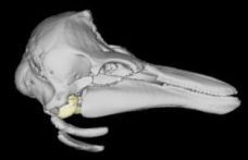
SSD-reconstruction from side elevation of an harbor porpoise skull. The light yellow marked area shows the bulla of the right ear (Petro-Tympanicum).
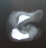
Left ear of a harbour porpoise in the decalcification phase (4. week). The bright white areas are calcified parts of the os petrosum (below) and os tympanicum (above). The darker white parts are decalcified collagenous areas. X-ray image was taken with a Unimat/Comet at 47 kV, 200 mA, 0.02 sec.