The incubation period of classical swine fever virus (CSF virus) in individual animals is about seven to ten days. Infection with low or moderately virulent strains that do not cause typical signs of disease result in longer high-risk periods (up to several weeks) facilitating unnoticed spread of CSF to other farms.
Classical swine fever (CSF) is characterized by extremely variable clinical signs and may be confused with many other diseases (e.g. African swine fever, porcine reproductive and respiratory syndrome, post-weaning multisystemic wasting syndrome, porcine dermatitis and nephropathy syndrome; erysipelas, salmonellosis, pasteurellosis, and other bacterial infections causing septicaemia; cumarin poisoning, purpura haemorragica.
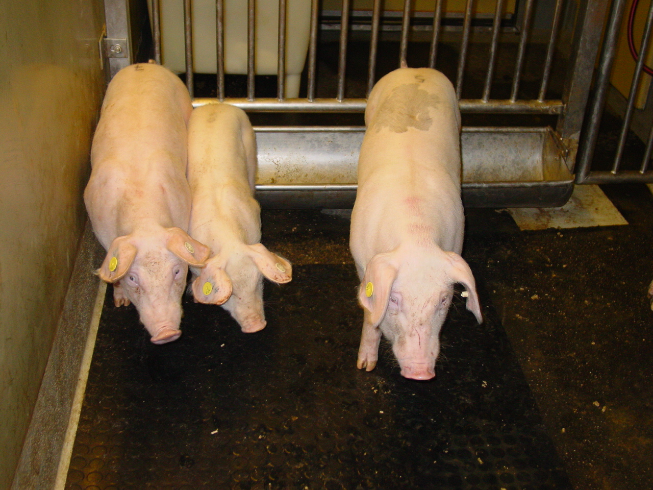
The virulence of the virus as well as the age of the animal influence the course of the disease. Losses in young animals are generally more severe than in adults.
Generally, three major clinical forms of CSF can be distinguished:
Acute form:
- Typically caused by highly virulent CSF virus strains, but also moderate strains can be involved
- Often observed in piglets up to 12 weeks
- First clinical signs may be anorexia, lethargy, fever (>40°C), conjunctivitis, swollen lymph nodes, constipation and/or diarrhoea. Neurological symptoms are frequent and can include, e.g. convulsions, uncoordinated movements and staggering gait.
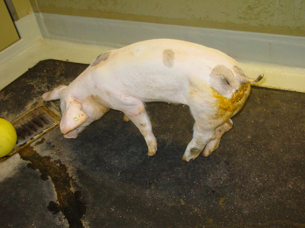
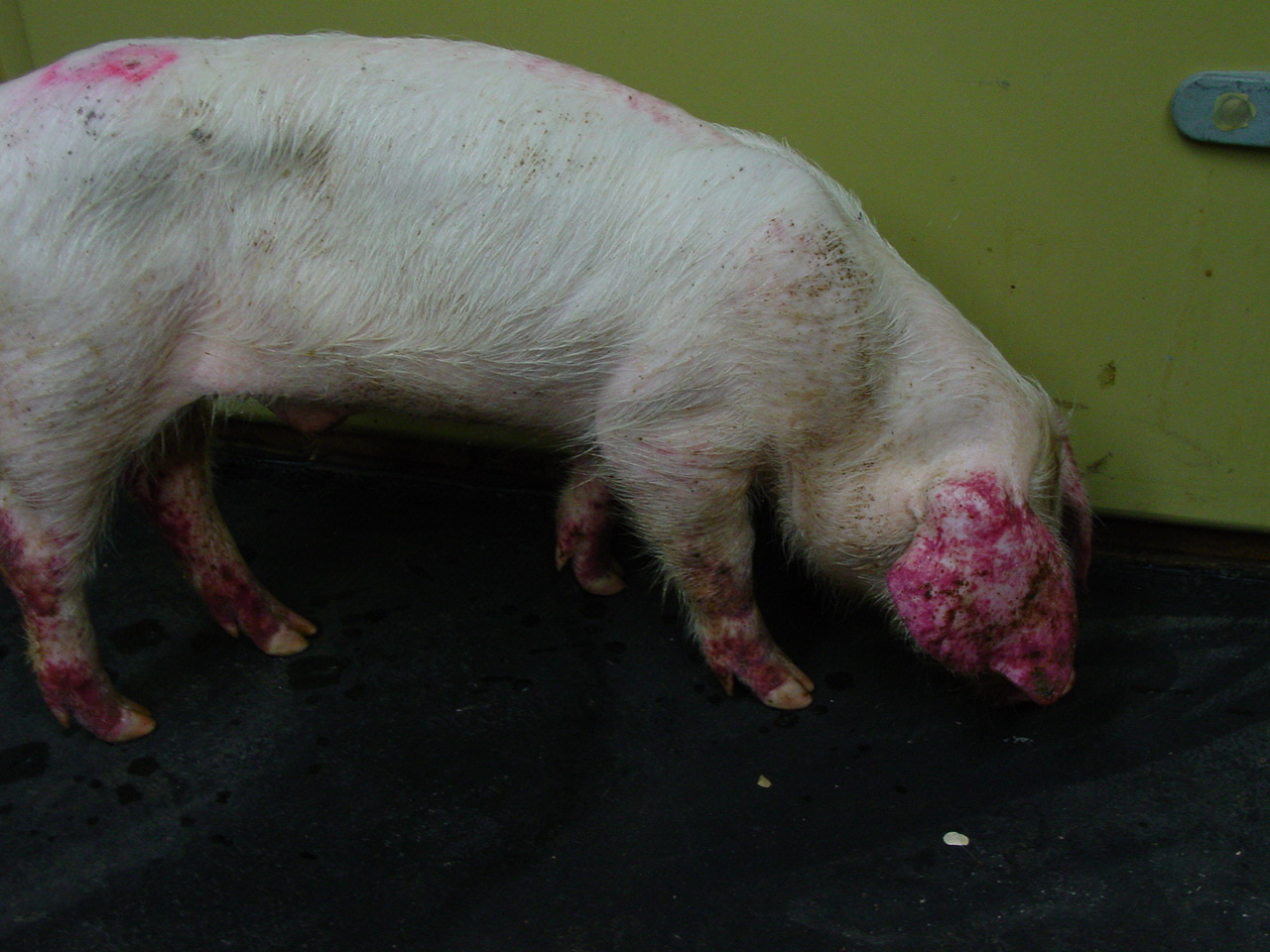
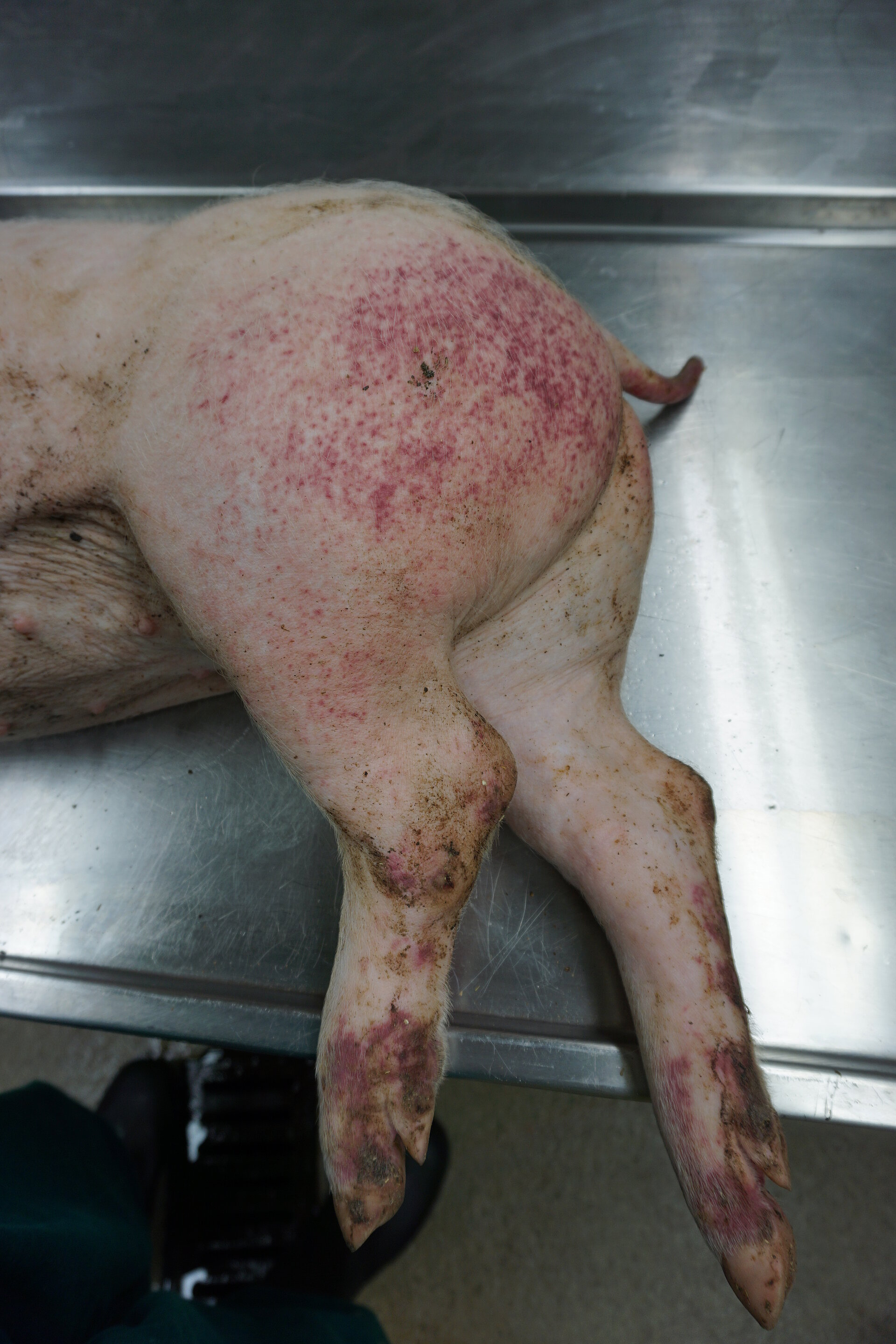
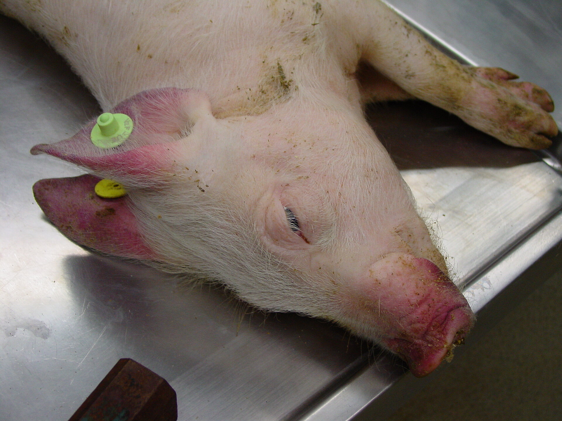
- Most characteristic feature is the hemorrhagic syndrome. About 2 weeks after infection, petechiae of the skin and mucosae, cyanosis of the ear, snout, tail, abdomen and medial side of the extremities might be detected.
- Severe leukopenia and immunosuppression can lead to enteric or respiratory secondary infections.
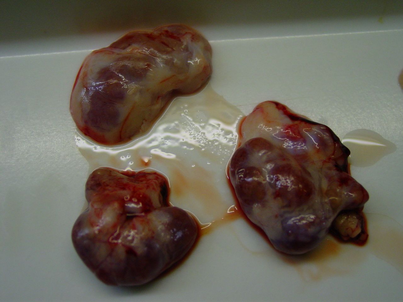
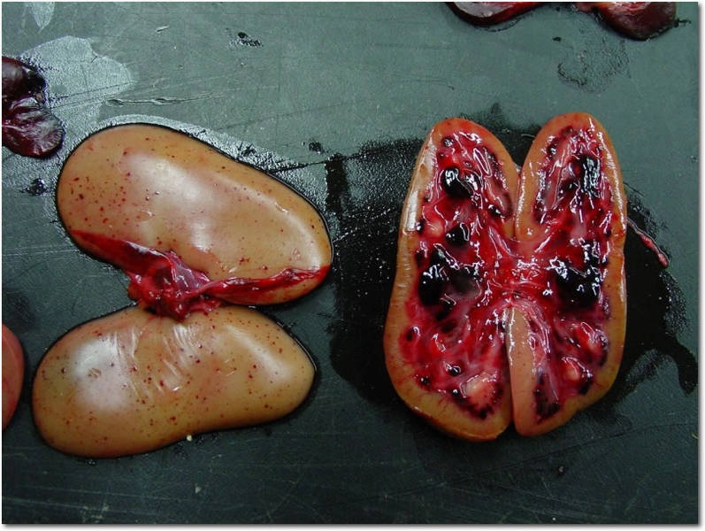
- Pathological changes include hemorrhages and/or petechial bleedings in tonsils, kidneys, spleen, serosae, lymph nodes, urine bladder and gastro-intestinal tract, as well as other organs.
- Lethality is close to 100% (between 10 – 20 days post infection)
- Recovery with production of antibodies does occur, most often in adult animals which do not display severe clinical signs. Antibodies against CSF virus are detectable from 2-3 weeks post infection onwards.
- CSF virus is shed from all mucosal surfaces resulting in its presence in saliva, urine and faeces from the onset of clinical signs until death. CSF virus can also be shed via semen.
Chronic form:
- Occurs mostly in young infected pigs that do not develop an effective immune response against CSFV infection. The immune response is insufficient to eliminate the virus.
- Initial clinical signs are similar to the acute form. Later, predominately non-specific signs are present, intermittent fever, chronic enteritis and wasting. The typical haemorrhages of the skin are missing.
- Animals can survive 2 – 3 months and shed virus from the onset of clinical signs constantly until death. Antibodies may be temporarily detected in serum samples.
- Pathological changes are less typical, especially concerning the lack of haemorrhages on organs and serosae. Thymus atrophy, ulcerative and necrotic lesions in the ileum, the ileocaecal valve and the rectum are common in pigs with chronic diarrhoea.
Prenatal form and late onset:
- CSFV can cross the placenta at any stage of pregnancy.
- Depending on the time of gestation and virus virulence, infection during early pregnancy can result in abortions and stillbirths, mummification and malformations or in birth of weak or apparently healthy piglets persistently infected with CSFV. All this leads to a reduction of the fertility index in the holding.
- Generation of persistently infected offspring occurs often via transplacental transmission of lower virulent CSFV strains.
- Persistently infected piglets can survive for several months without inducing antibody response against CSFV and play a crucial role in spreading the virus within the population while remaining undetected in serological tests.
- Piglets may appear clinical healthy at birth, or show unspecific signs, e.g. show poor growth, wasting or petechial skin haemorrhages or occasionally congenital tremor (“late onset of CSF”).
© TiHo Hannover, Institut für Virologie
© TiHo Hannover, Institut für Virologie
© TiHo Hannover, Institut für Virologie
© TiHo Hannover, Institut für Virologie

© TiHo Hannover, Institut für Virologie
© TiHo Hannover, Institut für Virologie
© TiHo Hannover, Institut für Virologie
© TiHo Hannover, Institut für Virologie