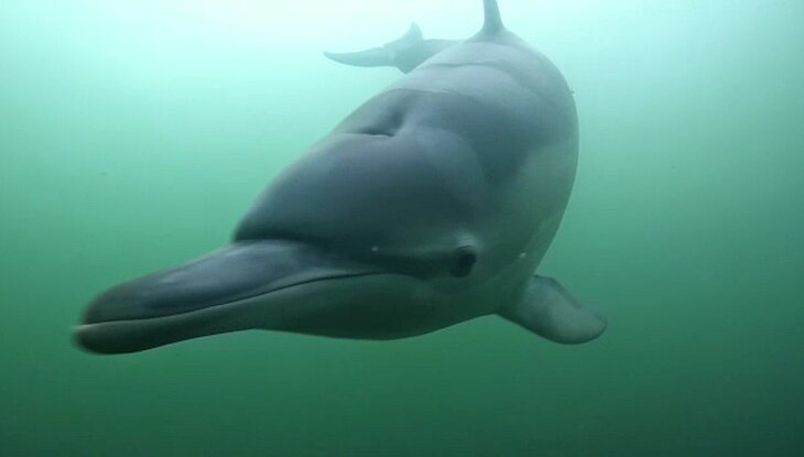
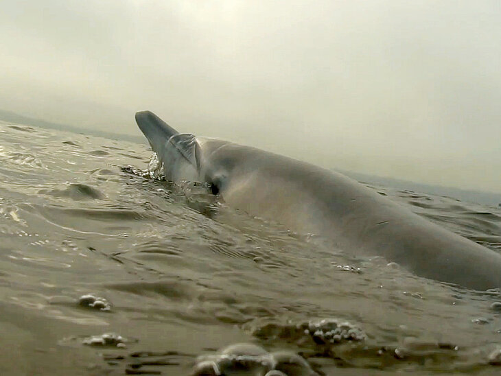
The Institute for Terrestrial and Aquatic Wildlife Research (ITAW) of the University of Veterinary Medicine Hannover (TiHo) has performed an autopsy on the common dolphin (Delphinus delphis) that had been in Eckernförde Bay for several months. The animal was found dead on the seabed at the end of January this year, recovered by divers and examined at the ITAW by veterinarians specializing in marine mammals. The comprehensive autopsy as well as further histological, bacteriological, virological and parasitological examinations as well as imaging methods of the ears were carried out in part also with partner specialist institutions.
The autopsy results
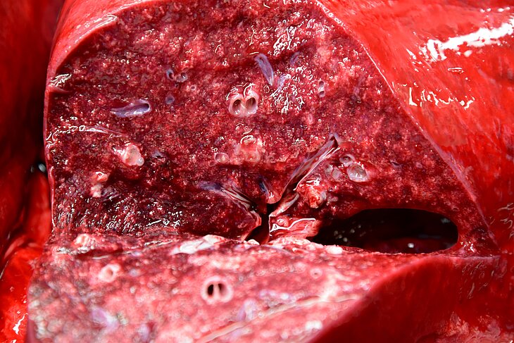
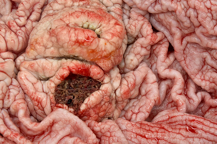
The female animal was about six years old and had not yet reached sexual maturity. The autopsy revealed abnormalities in the lungs of the young female dolphin. The tissue was infested with parasites and showed severe pneumonia. As a result, calcifications, connective tissue proliferation (fibrosis) and other severe changes in the tissue had occurred. In the stomach, the veterinarians also found parasites and two large and deep gastric ulcers. Significant disease was also indicated by the reactions of several lymph nodes in the lungs, intestines, stomach and neck, which, like in humans, are part of the dolphin's immune system.
Irregular, superficial lightening of the skin was visible throughout the animal's body. Last year, when the dolphin was still swimming in Eckernförde Bay, a skin disease had already been observed, which subsided after a few months and may have been caused by a smallpox infection. Investigations showed that the animal had recovered well from these changes.
A wound on the animal's head that had been noticed during recovery could be classified, after close examination and histological studies, as a superficial abrasion of the skin that the animal had contracted prior to death but was not relevant to the dolphin's death.
Antibody testing provided evidence that the animal had undergone a bacterial infection with the glanders pathogen Erysipelothrix rhusiopathiae. The bacterium is also transmissible to humans in close contact. At the time of the autopsy, the pathogen could no longer be detected. Testing for viral pathogens also did not yield positive results.
Since dolphins rely on their hearing to orient themselves, communicate with conspecifics or search for food, the veterinarians examined the dolphin in detail for acoustic impairments during the autopsy. Among other things, they used high-resolution imaging techniques to precisely assess whether the ears were damaged. In doing so, they were able to rule out the possibility that the dolphin had a fracture of the ear bones, for example.
The extensive examinations showed that despite the unchanged behavior still observed on the days before its death, the animal suffered from considerable health restrictions, especially of the lungs, which most likely led to a natural death of the young dolphin.
A video of the recovery of the dead dolphin can be found here: cloud.tiho-hannover.de/index.php/s/TJ7GFaTXm8zwXDr
Footage of the recovery of the dolphin. The divers used an airlift bag to bring the animal to the sea surface, where it was then lifted onto a boat. The video was taken by Jens Höhner.
Video of an autopsy
Using the example of a harbor porpoise, ITAW director Professor Dr. Ursula Siebert explains how the autopsy of an animal basically proceeds.
https://www.youtube.com/watch?v=5-uA0nWdQV0
Contact
Prof. Prof. h. c. Dr. Ursula Siebert
University of Veterinary Medicine Hannover
Institute for Terrestrial and Aquatic Wildlife Research
Phone: +49 511 856-8158
ursula.siebert@tiho-hannover.de


