3R SMART
Project Description:
Directive 2010/63/EU obliges a more consistent implementation of the 3Rs principle in the use of animals for scientific purposes, with the consequence of increased research activities in this field. The Foundation of the University of Veterinary Medicine Hannover and the Philipps University Marburg want to support these 3R research activities in a joint effort through a pilot project to establish an open access training platform for 3R methods and to make it more transparent and visible. The 3R training platform for Methodological Approaches to Reduce Animal Testing (3R-SMART) is competence-oriented and adapted to the needs of scientists, students, as well as technical staff at universities, public research institutions and in industry. To ensure broad acceptance of 3R-SMART among this target group, the EU Center for Alternatives to Animal Testing, the German Center for the Protection of Laboratory Animals (Bf3R) at the BfR, the Society for Laboratory Animal Science (GV-SOLAS), the Universities of Marburg and Konstanz, the Free University of Berlin with the Berlin-Brandenburg research platform BB3R, the Paul Ehrlich Institute, and BASF are involved in its content design.
To ensure high visibility from the beginning, 3R-SMART will be integrated into the established training platform for laboratory animal science (LAS interactive), which is operated by the Philipps University of Marburg. In addition, this concept allows the proven LAS interactive structures and content to be used for 3R-SMART. LAS interactive is focused on application-oriented online contributions to the refinement of animal experiments. These contributions will initially form the basis for the refinement module of the 3R-SMART, so that the development of the 3R-SMART can focus on training on methodological "replacement" and "reduction" approaches to animal testing.
In accordance with EU standards, the teaching of knowledge on the 3Rs principle is essential in courses to acquire expertise in laboratory animal science. Therefore, the 3R-SMART will also be used to develop a curriculum that allows to prove these 3R competences by an online examination. This curriculum will be developed in close cooperation with GV-SOLAS, which certifies laboratory animal science courses according to European standards.
After the project duration of three years, it is intended to expand the degree of dissemination of the 3R-SMART to the European level. For this purpose, the 3R-SMART is to be included in the EU platform "Education and Training in Laboratory Animal Science" (ETPLAS). To support this sustainability strategy, the 3R-SMART will be comprehensively evaluated twice during the project period. In this way, the proposed project can make a significant and sustainable contribution to a wider use of 3R methods and a correspondingly reduced use of laboratory animals.
Funding:
3R-SMART, German Federal Ministry of Education and Research (BMBF)
Contact:
Prof. Dr. Bernhard Hiebl (bernhard.hiebl(at)tiho-hannover.de; +49 511-856-8958)
Institute for Animal Hygiene, Animal Welfare and Farm Animal Behaviour
Altered protein and lipid transport in intestinal epithelial cells
Studies on altered protein and lipid transport in stable cell lines after contact with toxic substances as a cause of pathological conditions
Methods:
- polar intestinal epithelial cells
Project Description:
For the development of an effective therapy for inflammatory bowel diseases in mammals, triggered by genetic factors or by the ingestion of toxic substances via the diet, a precise knowledge of the biochemical and physiological processes in the affected epithelial cells of the inflamed intestinal segment is essential. An understanding of the epithelial cells in the pathological state is as important as, for example, that of the morphology of the cell membrane and its components in the healthy intestine. The CaCo-2 cell model will be used to characterize the basic interaction possibilities between toxic substances from food and epithelial cells. Attention will be paid to cause-effect analyses of the adhesion of the substances, their endocytosis, and their trafficking within epithelial cells. The human colorectal cell line CaCo-2 has long been a scientifically accepted model for intestinal cells, with many properties of membrane topology, cell biochemistry, and transport that correspond to those of physiologically inconspicuous intestinal mucosal cells. Special cultivation in snap-well inserts allows the apical/intestinal lumen uptake of substances to be simulated and the basolateral/tissue release of metabolites to be analyzed. In addition, the treated cells are examined biochemically and cell biologically for pathological changes.
Contact:
Prof. Dr. Hassan Y. Naim (hassan.naim(at)tiho-hannover.de; +49 511-953-8787)
Institute for Biochemistry
Bacterial infections in humans and animals
In vitro and in vivo characterization of the innate immune response against bacterial infections in humans and animals
Methoden:
- Primary neutrophils (human, porcine, bovine, equine, mouse)
- Primary lung epithelial cells (human, mouse, pig)
- Choroid plexus epithelial cells (human, pig)
- Intestinal epithelial cells (Human)
- Primary mast cells (human, mouse)
Increased complexity, which should more closely resemble the in vivo situation, is enabled by physiologically relevant oxygen conditions and by 3D co-cultivation of epithelial cells and neutrophils. A hypoxia glove box is available for defined hypoxic conditions.
Project Description:
The increased incidence of resistant bacteria worldwide limits the efficiency of antibiotic-based treatment concepts. New promising therapeutic approaches are needed, such as enhancing host defenses by stimulating the immune system to support conventional antibiotic-based therapy. However, the complex host-pathogen interactions remain poorly elucidated. Detailed knowledge is required to apply therapeutic strategies based on innate immune defense. However, due to lack of complexity, incorrect cell differentiation status, and lack of physiological conditions, in vitro systems do not always accurately simulate the in vivo situation during infection or inflammation. Results from conventional in vitro experiments investigating host-pathogen interaction or therapeutic approaches often contradict data from in vivo studies and are therefore difficult to extrapolate. Non-animal test systems for both infection and interaction studies and drug screenings are only a real alternative if the results obtained can be reliably transferred to an in vivo situation.
Therefore, our goal is to establish improved in vitro model systems that reliably simulate the innate immune response against bacterial pathogens. Increased complexity of the model system will allow the in vitro system to approximate the in vivo situation, thereby helping to reduce the number of experimental animals.
Funding:
R2N (2017-2021) funded by the Ministry of Science and Culture of Lower Saxony
Contact:
Prof. Dr. Maren von Köckritz-Blickwede, (Maren.von.Koeckritz-Blickwede(at)tiho-hannover.de +49 511-856-8787)
Institute for Biochemistry and Research Center for Emerging Infections and Zoonoses (RIZ)
Biocompatibility of nanoparticles
In vitro test battery for testing the biocompatibility of nanoparticles
Methods:
- Cell culture of murine dendritic cells, macrophages and osteoblasts to study the biocompatibility of implants, nanoparticles and other test substances
- Cell migration assays
- Viability assays
- Proliferation assays
- Measurement of cytokine release (cell culture and tissue sections)
Project Decription:
In a DFG project, magnetic nanoparticles are being developed for use as carriers for active substances. In order to test the release of active substances and the biocompatibility of the nanoparticles, meaningful in vitro model systems are being developed. In addition to work on cell cultures, the isolated perfused bovine udder (ex vivo model) is established in our own working group for this purpose. The ultimate goal of the project is to establish an in vitro test battery for biocompatibility testing, which will allow the selection of nanoparticles suitable for in vivo use.
Funding:
DFG
Contact:
Dr. Jessica Meißner (jessica.meissner(at)tiho-hannover.de; +49 511-953-8723)
Institute for Pharmacology, Toxicology and Pharmacy
Canine and murine slice cultures of the central nervous system
Organotypic canine and murine slice cultures as an in vitro model for studying glial responses and interactions in neurotraumatic, -infectious, and -degenerative diseases of the central nervous system
Methods:
Organotypic slice cultures of the central nervous system are to be regarded as an intermediate model between animal experiments in vivo and dissociative cultures in vitro and represent a suitable model to validate findings in single cell cultures and to reduce the use of animal experiments. In sectional cultures, the organotypic tissue structure is largely preserved in contrast to dissociative cell culture, so that conditions in vivo can be simulated reductionistically and complex interactions of glial cells can be studied without the influence of peripheral inflammatory cells. Slice cultures also offer the possibility of targeted, for example pharmacological, manipulation of culture conditions and easy visualization of resulting effects. Preliminary screening of potential therapeutic candidates is thus possible without the use of animal experiments.
In our group, methods have been established that allow the preparation of slice cultures post-mortem. The removal of brain or spinal cord is performed under sterile conditions in the context of an autopsy of the animals. Using a tissue chopper or alternatively a vibratome, 350 µm wide tissue slices are prepared, which are then cultured on suitable membrane inserts with the addition of a suitable cell culture medium. The slice cultures are then formalin-fixed after a specified cultivation time and subsequently examined histologically, immunohistologically and/or molecularly. The preparation of numerous slice cultures from one animal allows the simultaneous performance of several experiments and is associated with a significant reduction in the number of animals that would be necessary for in vivo studies.
Project Description:
Future research projects in our group are based on already published observations in canine and murine slice cultures. Since the slice preparation itself simulates trauma, temporal changes in the tissue can be considered as a model for processes after neurotrauma. Thus, organotypic slice cultures from adult canine spinal cord have already been established as a complementary model for processes in the injured canine spinal cord, which is characterized by a distinct response of phagocytotically active microglia/macrophages comparable to the situation in vivo (Spitzbarth et al., 2011; Bock et al., 2013). Furthermore, in additional studies, we demonstrated that culturing slices of the canine olfactory bulb and brainstem, as well as culturing different localizations of adult mouse brain, is associated with spontaneous proliferation of p75 neurotrophin receptor (p75NTR)-positive, potentially regeneration-promoting Schwann cell-like glial cells (Imbschweiler et al., 2012; Spitzbarth et al., 2015; Kegler et al., 2015). Furthermore, preliminary studies have shown that slice cultures of adult canine brain can be infected with canine distemper virus, making this in vitro model also suitable to study certain aspects of canine distemper virus leukoencephalitis in an animal-independent manner (Lempp et al., 2014).
Future research will further investigate the interaction between potentially regenerating Schwann cell-like glial cells with microglia/macrophages to identify potential trigger factors that promote proliferation of p75NTR-positive glia. Pharmaceuticals may also be used for this purpose to manipulate culture conditions (e.g., minocycline to suppress microglia/macrophage activation). Similarly, canine brain slice cultures will be infected with canine distemper virus, target cells of infection will be characterized, and findings will be comparatively evaluated with uninfected control slices to gain insight into the pathogenesis of glial changes in canine distemper virus leukoencephalitis.
Contact:
Prof. Dr. W. Baumgärtner, Ph.D. (wolfgang.baumgaertner(at)tiho-hannover.de; Tel.: +49 511-953 8620)
Prof. Dr. A. Beineke (andreas.beineke(at)tiho-hannover.de; Tel.: +49 511-953 8640)
Institute for Pathology
Cell culture system for the investigation of hepatitis E virus (HEV) infection
Establishment of a reverse genetic system to study molecular mechanisms of hepatitis E virus (HEV) infection
Methods:
- establishment of a cell culture model for hepatitis E virus (HEV, genotype 3)
- cloning of the whole genome of HEV into a bacterial vector
- establishment of a reverse genetic system for HEV
Project Description:
Since the introduction of mandatory reporting of human hepatitis E disease in 2001, the number of reported cases has been steadily increasing in Germany. In Europe, HEV genotype 3 in particular leads to chronic manifestations of disease in immunocompromised patients. In addition to deer species and wild boar, domestic swine have been identified as the reservoir host in Europe. Many aspects of pathogenesis and epidemiology, including the mode of transmission from animals to humans, are poorly understood. On the one hand, the mechanisms of the different virulence expression in the reservoir host pig and in humans are unclear. Furthermore, no studies on cellular susceptibility and the cellular receptor are available. Furthermore, large parts of the multiplication cycle of HEV have not yet been investigated. This is mainly due to the fact that only very few selected HEV strains can be cultured in vitro and established cell culture models are not yet available to study the molecular basis of HEV replication. In contrast, HEV infection is well characterized in animal models (mainly swine).
An important goal of the project is the cloning of the whole genome of HEV into a corresponding vector, in order to subsequently carry out various genetic modifications at the DNA level. After transcription into RNA, this is introduced into susceptible cells by electroporation. In this way, basic mechanisms of virus replication and release can be studied in vitro. The project includes the development of a cloning strategy and its implementation, the selection of a suitable cell culture system and the generation of infectious virus particles. The establishment of this system will allow detailed studies on different aspects of HEV infection in cell culture, which so far cannot be investigated or can only be investigated in animal experiments, and will thus contribute to the reduction of animal experiments.
Contact:
PD Christine Bächlein, Ph.D. (christine.baechlein(at)tiho-hannover.de; Tel.: +49511-953-8845),
Prof. Dr. Paul Becher (paul.becher(at)tiho-hannover.de; Tel.: +49 511-953-8840)
Institute for Virology
Cell differentiation in intestinal organoids
Development of antibiotic resistance
FERTHIK II
Teaching veterinary clinical skills and implementing ethics in veterinary medicine
Methods:
- Practical exercises on models
- Development of models
- Supervised small group peer teaching
- Learning through video material
- Reflection in terms of animal welfare and ethics
Project Description:
The project "Teaching veterinary clinical skills and implementation of ethics in veterinary medicine (FERTHIK II)" is a follow-up project of "FERTHIK - Teaching veterinary clinical skills under special recognition of ethical aspects". In the course of these projects, the Clinical Skills Lab (CSL) was established at the TiHo and the range of learning stations was continuously expanded. In the CSL, learning and practicing clinical skills on models is possible for veterinary students of all semesters, with the effect that the exercises on live animals are not further extended. FERTHIK aims to improve teaching in the area of practical skills of students in dealing with pets and farm animals, taking into account animal welfare and ethical issues (skills and attitudes), thus increasing the practical relevance of veterinary studies. Within the project, simulators and models are developed in the Skills Lab in collaboration with the clinics and institutes of the TiHo. As a further component of the learning stations and models in the CSL, in addition to practicing the practical activities, questions of animal welfare and animal ethics are discussed, which are related to practical activities and which a veterinarian is confronted with. By deliberate practice of practical skills on models in a "safe" environment, students develop a routine in performing a veterinary skill before the first intervention on a live animal. The students also have the opportunity to perform so-called "day-one competencies" several times on the simulators and to develop their own self-confidence through repeated practice in order to be well prepared and competent as a novice in the everyday life of a veterinarian, which also benefits animal welfare in practice. The clinical skills that can be practiced at the learning stations are recorded and the video material for preparation and follow-up is made available on the YouTube channel TiHoVideos.
Funding:
FERTHIK II, Germany Federal Ministry of Education and Research (Federal and State programme: Teaching Quality Pact)
Contact:
Prof. Dr. Andrea Tipold (Andrea.Tipold(at)tiho-hannover.de; +49 511-953-6411)
Small Animal Clinic
Dr. Elisabeth Schaper (Elisabeth.Schaper(at)tiho-hannover.de; +49 511-953-8036)
Center for E-Learning, Didactics and Educational Research (ZELDA)
Dr. Sandra Wissing (Sandra.Wissung(at)tiho-hannover.de; +49 511-856-8360)
Center for E-Learning, Didactics and Educational Research (ZELDA)
Prof. Dr. Peter Kunzmann (Peter.Kunzmann(at)tiho-hannover.de; +49 511-856-8959)
Institute for Animal Hygiene, Animal Welfare and Farm Animal Behaviour
Glycan array platform
Glycan array platform for in vitro prescreening for carbohydrate-based adjuvants and immunomodulators
Methods:
- Glycan and lectin arrays
- In vitro co-cultures of antigen-presenting cells and T cell receptor transgenic T cells
Project Description:
Testing of adjuvants and immunomodulators in vivo requires a large number of experimental animals, so appropriate in vitro prescreening is urgently needed to minimize animal numbers for in vivo studies. This project will develop a new platform to screen for carbohydrate-based adjuvants and immunomodulators. Immobilized synthetic glycans or glycans isolated from pathogens will be immobilized on microarray chips and tested for binding to C-type lectin receptors (CLRs) of innate immunity. Myeloid CLRs are carbohydrate-binding proteins (known as lectins) that are expressed primarily by antigen-presenting cells and can recognize sugar structures on pathogens and thereby initiate adaptive immune responses. Targeting CLRs is a promising strategy to enhance immune responses against vaccine antigens or even to specifically modulate immune responses (Lepenies et al., Adv. Drug. Deliv. Rev. 2013, 65, 1271-81). In preliminary work, a library of CLR-Fc fusion proteins was generated in which the extracellular portion of each CLR was fused to the Fc fragment of human IgG1 molecules (Maglinao et al., J. Control. Release 2014,175, 36-42). The generated CLR-Fc library now includes all immunologically relevant murine CLRs and will be extended to human and ovine CLRs in this project. Carbohydrates identified as CLR ligands by glycan array are subsequently coupled to model antigens and tested for their immunostimulatory or modulatory properties in vitro in co-cultures of dendritic cells and T cell receptor transgenic T cells (Brzezicka et al., ACS Chem. Biol. 2016, 11, 2347-56). This in vitro prescreening allows pre-selection of suitable glycans as candidates for new adjuvants and immunomodulators, thus reducing the number of experimental animals for immunization studies.
Contact:
Prof. Dr. Bernd Lepenies (bernd.lepenies(at)tiho-hannover.de +49 511-953-6135)
Marie-Kristin Raulf (Marie-Kristin.Raulf(at)tiho-hannover.de +49 511-953-6124)
Institute Immunology & Research Center for Emerging Infections and Zoonoses
Innervated Skin Model
Development of a human innervated skin model for the identification of skin sensitizing substances
Methods:
- Differentiation of human induced pluripotent stem cells (hiPSCs) into sensory neurons
- Differentiation of hiPSCs into Schwann cells
- Co-culture of the involved cells (sensory neurons, Schwann cells, fibroblasts, keratinocytes) in a hydrated matrix
- Quantification of neurite outgrowth in a skin model
Project Description:
Products of daily life such as cosmetics, clothing or medicines come into contact with the skin. Sensitization of the skin, triggered by contact allergens, manifests itself as an unpleasant allergic contact dermatitis accompanied by itching. In Europe, about a quarter of the population is affected. Manufacturers must ensure that substances that come into contact with the skin do not trigger contact allergy. The identification of skin sensitizing substances is based on an Adverse Outcome Pathway (AOP) with certain key events that can be investigated via in vitro activation of keratinocytes, dendritic cells and in vivo activation of T cells. The so-called adverse outcome, i.e. allergic contact dermatitis, can so far only be studied via poorly representative animal experiments.
In this project, a 3D skin model containing sensory neurons and Schwann cells derived from human iPSCs will be developed to quantify the increased neurite growth in vitro that corresponds to pruritus, a key symptom of allergic contact dermatitis. This assay could complement the existing AOP and allow to significantly reduce the gap between in vitro experiments and in vivo induced allergic contact dermatitis
Contact:
Prof. Bettina Seeger, Ph.D. (bettina.seeger(at)tiho-hannover.de; +49 511-856-7602)
Maren Schenke, Ph.D. (maren.schenke(at)tiho-hannover.de; +49 511-856-7603)
Institute for Food Quality and Food Safety
Insect embryos for developmental neurotoxicity testing
An intact insect embryo as a test system for safety toxicology testing for developmental neurotoxicity
Methods:
- Serum-free embryo culture
- Immunofluorescence
- Neuroanatomy
Project Description:
Developmental neurotoxicity (DNT) describes the functional and morphological effects on the developing nervous system of the offspring that can occur as a result of exposure to contaminants during pregnancy and early postnatal development. The in vivo tests currently approved for testing substances for DNT involve very high animal consumption and are laborious and stressful for the test animals. In this project, a simple evertebrate test system was developed that can reflect the complexity of the pathfinding processes involved in embryonic brain development. This experimental system captures, as a functional endpoint following chemical exposure, the perturbations of axonal wayfinding of identified pioneer axons in the leg bud of locust embryos as they move into the central nervous system. An essential aspect of brain development is precise linkage between neurons. Following chemical exposure, the assay system detects disruptions in neuronal circuitry from identified pioneer axons to the central nervous system. The outgrowing cell processes of these pioneer neurons make their way to the central nervous system along semaphorin signpost molecules. The cellular mechanisms of this navigation of neuronal projections have not been altered between invertebrates and the mammalian cortex during evolution. Therefore, sensitivity to hazardous chemicals in the insect embryo assay also allows conclusions to be drawn about developmental neurotoxicity in humans. Another goal of this project was the 3D acquisition of pioneer morphology using Scanning Laser Optical Tomography (SLOT). For this purpose, fixed embryonic stages were treated with different histological clearing methods. Our project partners at the Laser Zentrum Hannover (LZH) generated the raw optical tomographic data and calculated the corresponding 3D projections. In collaboration with the LZH, it was shown that the established developmental neurotoxic compounds methylmercury and sodium arsenite for axonal navigation were also identified as DNT-positive by the SLOT procedure. The SLOT procedure, in combination with machine vision segmentation techniques, could allow more accurate classification of pathfinding errors.
https://www.youtube.com/watch?v=SrdjplVsWto
Contact:
Prof. Dr. Gerd Bicker (gerd.bicker.iR@tiho-hannover.de; +49 511-856-7765)
PD Dr. Michael Stern (michael.stern@tiho-hannover.de; +49 511-856-7767)
Institute for Physiology and Cell Biology
Intestinal organoids and intestinal epithelial cells for the study of intestinal diseases
Development of an in vitro model to study the pathogenesis mechanisms of zoonotic pathogen-induced intestinal diseases
Methods:
- Coculture of human enterocytes and goblet cells
- Cultivation of intestinal organoids from pigs
- Analysis after treatment with bacterial toxins with
- Ussing chamber
- RT-qPCR
- Western blot
- immunofluorescence
Project Description:
The intestine plays a crucial role for many diseases transmissible from animals to humans (so-called zoonoses). Both in the establishment of an infection, as well as for the excretion of pathogens and the potentially associated spread of infection. While various cell culture-based in vitro systems are available for the study of disease mechanisms in mice, rats, and humans, the study of such processes in farm animals (e.g., pigs, cattle, or poultry) still requires the use of primary cell or organ cultures taken directly from the animal, or even the use of animals. For this reason, new approaches to develop in vitro models to study underlying molecular mechanisms of infections in farm animals are of great interest, especially in light of the fact that farm animals are often carriers or vectors of zoonotic pathogens. The purpose of the present project is therefore to develop a model of the intestine by combining for the first time the so-called "colon simulation technique" (Cositec) with the "Ussing chamber technology". Intact intestinal mucosa from farm animals is clamped in Ussing chambers and incubated simultaneously with the contents of the Cositec system, so that a confrontation of the intestinal epithelium with the physiological intestinal contents can take place. The combination of these two well-established methods thus makes it possible to design an in vitro model whose conditions correspond as closely as possible to in vivo conditions. In a first step, the vitality and functionality of the intestinal epithelium in this system are assessed by morphological, biochemical and functional analyses (e.g. by histology, PCR or nutrient uptake). Of great importance is that the intestinal tissue used for all analyses does not originate from laboratory animals, but is always obtained from conventional slaughterhouses. This means that the use of laboratory animals can be dispensed with completely. In a subsequent step, the intestinal epithelium is to be incubated with zoonotic pathogens (e.g. enterotoxic and enteropathogenic E. coli) in order to be able to investigate infection mechanisms of these pathogens and their effects on the intestine in more detail. If this new system were to work, it would have a variety of uses apart from the study of infectious diseases. These include, for example, physiological, toxicological or even pharmacological questions. Furthermore, cell cultures of the intestine could also be used in this model, which would make the use of animal material largely superfluous. Overall, a successful establishment of this system would result in a significant reduction in the number of laboratory animals.
Funding:
R2N, Ministry of Science and Culture of Lower Saxony, 2017-2021
Contact:
Prof. Dr. Gerhard Breves (gerhard.breves(at)tiho-hannover.de, +49 511-856-7271)
Institute for Physiology and Cell Biology
Prof. Bettina Seeger, Ph.D. (bettina.seeger(at)tiho-hannover.de; +49 511-856-7602)
Institute for Food Quality and Food Safety
MoNLight-BoNT assay
MoNLight-BoNT assay - Development of a serotype-independent botulinum neurotoxin activity assay for batch testing
Methods:
- Differentiation of human induced pluripotent stem cells (iPSCs) into motor neurons
- RT-qPCR
- Western blot
- luminescence assay
Project Description:
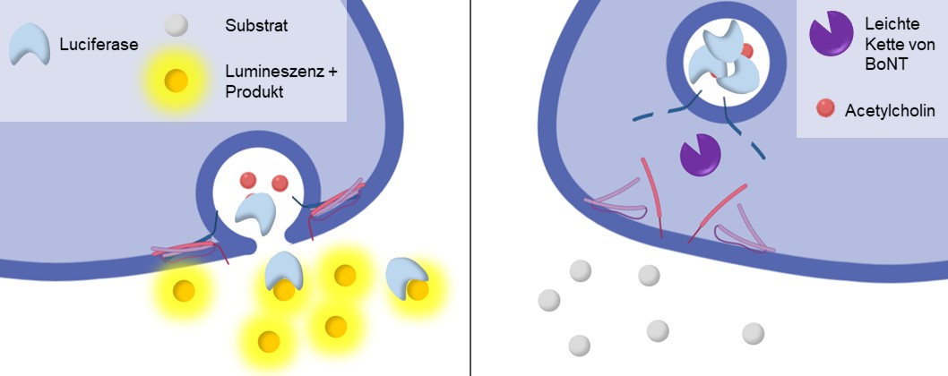
Botulinum neurotoxins (BoNTs) are used to treat a range of neuromuscular dysfunctions and in aesthetic medicine as therapeutic agents. Each batch of pharmacological preparations of these highly potent neurotoxins, produced by Clostridium botulinum bacterial strains, must be tested for activity. Although alternatives exist, their activity is still determined to a large extent by the mouse lethality assay. Replacement methods approved to date are applicable only to single compounds, where highly specific neoepitope antibodies are used to detect the proteolytic activity of the small subunit of BoNTs. For batch testing of BoNTs, 400,000 mice are still used annually within Europe alone (as of 2018). New products tested exclusively in animals are no longer accepted by the European Medicines Agency (EMA). More than 40 subtypes and mosaic variants are now known in addition to BoNT serotypes A-H. Pharmacological use of diverse subtypes and new products is likely in the future, as resistance to individual products may emerge. A serotype-independent, animal-free assay, which can be used for activity testing of BoNT-containing pharmacological products across the board, does not yet exist but is urgently needed.
The MoNLight-BoNT assay (Motor Neuron Light-BoNT Assay) is based on motor neurons (MNs) differentiated from human induced pluripotent stem cells (iPSCs). The activity of the pharmacological BoNT products is inversely proportional to the release of a reporter enzyme (luciferase) from the neurotransmitter-containing vesicles of MNs. The assay is serotype-independent and would therefore be a suitable replacement method to fully replace the mouse lethality assay associated with batch testing (Replace in the context of the 3Rs).
Funding:
German Ministry of Education and Research (BMBF; FKZ 031L0132B, 2017-2020)
Contact:
Prof. Bettina Seeger, PhD (bettina.seeger(at)tiho-hannover.de; +49 511-856-7602)
Maren Schenke, Ph.D. (maren.schenke(at)tiho-hannover.de +49 511-856-7603)
Institute for Food Quality and Food Safety
Network meta-analyses on omics studies
Network meta-analyses for indirect inference from published omics studies
Methods:
- Knowledge extraction from published studies and data
- Avoidance of animal testing through indirect inference from existing results
- Merging of study results with methods of Data Science
- Methods for simulating high-dimensional data structures
Project Description:
Indirect group comparisons can be made from existing study results using network meta-analyses, i.e. group comparisons that have not yet been investigated in the original studies. Thus, further animal experiments can be avoided. This project will investigate how network meta-analysis can be applied to high-dimensional data from "omics" experiments (e.g., transcriptome, proteome, or metabolome profiles). Research will also be conducted on how new knowledge can be extracted from already public experimental omics data using pattern recognition methods.
In addition, methods are being developed to simulate high-dimensional data in general. These methods can help to simulate transcriptome, proteome or metabolome studies on the computer.
Contact:
Prof. Dr. Klaus Jung (klaus.jung(at)tiho-hannover.de; +49 511-953-8878)
Institute for Animal Breeding and Genetics, AG Genomics and Bioinformatics of Infectious Diseases
Oxygen-dependent differentiation of mast cells
Precision cut bovine udder slices (PCBUS)
Precision cut bovine udder slices (PCBUS) as an in vitro model to characterize the early infection events of mastitis
Methods:
- Precision cut bovine udder slices (PCBUS)
Project Decription:
Precision cut slices of various organs are well established in pharmacological-toxicological research for the investigation of xenobiotic exposure, drug effects and toxicological issues.
In the research group, precision cut bovine udder slices were successfully established. These allow the investigation of the early infection process of mastitis in vitro by co-culturing with mastitis-relevant pathogens (e.g. Staphylococcus aureus). The complex tissue association of the precision sections allows a comprehensive characterization of tissue reactions to different pathogens. The next step will be to characterize in more detail the possibility of drug treatment in the early stages of infection.
Contact:
Dr. Jessica Meißner (jessica.meissner@tiho-hannover.de; +49 511-953-8723)
Institute for Pharmacology, Toxicology and Pharmacy
Precision cut equine lung slices
Precision cut equine lung slices as an in vitro model of obstructive airway disease for identification of potential therapeutic targets and drug characterization
Methods:
- Precision cut lung slices (PCLS)
Project Desciption:
Precision cut slices of various organs are well established in pharmacological-toxicological research for the investigation of xenobiotic exposure, drug effects and toxicological issues.
In collaboration with Prof. Ohnesorge (Clinic for Horses), precision cut lung slices from horses have been successfully established, which allow to characterize pathomechanisms of chronic obstructive pulmonary disease in horses in more detail and to investigate possible treatment approaches in vitro. The intact tissue association allows the investigation of complex disease processes.
Contact:
Dr. Jessica Meißner (jessica.meissner@tiho-hannover.de; +49 511-953-8723)
Institute for Pharmacology, Toxicology and Pharmacy
Primary cell and organ cultures of poultry species
Epithelial cells as targets for infectious agents: primary cell and organ cultures as complements or substitutes to animal experiments.
Project Description:
At the Clinic for Poultry, zoonotic pathogens such as avian influenza virus (AIV) and Campylobacter spp. as well as poultry-specific pathogens such as avian metapneumovirus, Bordetella avium or Mycoplasma gallisepticum are investigated in different studies. Epithelial cells of the respiratory, intestinal or sexual tract are target cells for these pathogens. Investigations of pathogen-host interactions on these target cells, especially in the context of co-infections, would require very extensive animal experiments under in vivo conditions.
Partial goals of our research projects are to better understand the pathogen-host interactions on epithelial cells, to identify species-specific differences between the different farm poultry species and to find approaches to better control these different pathogens in the field. For this purpose, we have established primary cell and organ culture systems in our research group in order to be able to investigate pathogen-host interactions under well standardized conditions while avoiding or reducing animal experiments. These organ and cell cultures include systems representative of the laying gut, the trachea, and different sections of the intestinal tract. The cell and organ cultures can be obtained from different avian species, allowing investigation of pathogen pathogenesis in vitro and pathogen-host interactions between different species simultaneously under comparable laboratory conditions. The epithelial character of these cell and organ cultures is preserved for several days, and microscopic examination of the ciliary beat, for example, can be used as a parameter to detect pathogen exposure. Furthermore, techniques such as qRT-PCR are used to study innate immune responses after infection. The replication of the respective pathogens is detected by classical virological or bacteriological but also molecular biological methods.
Contact:
Prof. Dr. Silke Rautenschlein (silke.rautenschlein(at)tiho-hannover.de; +49 511-953-8779)
Dr. Arne Jung (arne.jung(at)tiho-hannover.de +49 511-953-8774)
Cllinic for Poultry
Regulatory mechanisms of P-glycoprotein on brain endothelial cells
Regulatory mechanisms of P-glycoprotein on brain endothelial cells of different species and new in vivo approaches to translation of in vitro findings
Methods:
- Use of in vitro BBB models to study drug transporter-mediated regulatory mechanisms
Project Description:
Given the pharmacoresistance of common brain diseases such as epilepsy or depression to current therapies and the potential importance of efflux transporters such as P-glycoprotein (Pgp) for resistance mechanisms, deciphering the regulatory mechanisms of Pgp at the blood-brain barrier (BBB) and searching for innovative therapeutic approaches, are of fundamental importance. To investigate rapid, on-demand regulatory mechanisms of the efflux transporter Pgp at the human BBB for drug action, the human brain capillary endothelial cell line hCMEC/D3 was transduced with MDR1-EGFP to visualize Pgp. Cell-cell Pgp transfer and intracellular Pgp-mediated substrate sequestration are studied in this in vitro BBB model. Sequestered drugs are released from the cell in the form of extracellular vesicles that attach to blood-facing cell membrane and are disposed of by interaction with blood cells. The significance of these new mechanisms will be investigated comparatively with other Pgp regulatory mechanisms in hCMEC/D3 cells, as well as brain endothelial cells of other species in vitro. In this context, the different in vitro BBB models will be characterized and optimized. Among others, the influence of the tight junction protein claudin-5 on barrier properties in the human hCMEC/D3 model will be investigated. By means of different ex vivo methods, the in vivo relevance of Pgp regulatory mechanisms will be evaluated. We expect our studies to characterize novel cross-species or -specific regulatory mechanisms of Pgp at the BBB and, thus, the possibility of pharmacological intervention in resistance mechanisms of brain diseases.
Contact:
Prof. Dr. Wolfgang Löscher (wolfgang. loescher(at)tiho-hannover.de; Tel. +49 511-953-8729)
Institute for Pharmacology, Toxicology and Pharmacy
SALMHEARTCELL
SALMHEARTCELL - Development and use of primary and permanent cell cultures from salmonid heart cells for replication and detection of piscine orthoreoviruses
Project Description:
The SALHEARTCELL project aims to develop new marine resources by growing primary cultures of salmonid cardiac cells (SalCPCs) and permanent cell cultures from the heart tissue of Atlantic salmon (Salmo salar), rainbow trout (Oncorhynchus mykiss) and brown trout (Salmo trutta) and using these cells in studies of emerging viruses that cause circulatory disorders in salmonids. This will be achieved through the expertise and collaboration of the partners Fraunhofer Research Institution for Marine Biotechnology and Cell Technology (EMB) and the Department of Fish Diseases and Fish Husbandry of the University of Veterinary Medicine Hannover (TiHo). The cultures and cell lines will be thoroughly characterized and used in experiments to replicate and detect piscine orthoreoviruses 1 and 3 (PRV-1 and PRV-3). These emerging pathogens in fish have recently been detected in Germany, posing an immediate threat to German aquatic marine resources. PRV-1 and PRV-3 infect cardiomyocytes and erythrocytes. Despite various attempts, the cultivation of these viruses in existing cell lines failed. This severely compromises disease diagnosis, virus research, and vaccine development that could mitigate the threat. SalCPCs contain spontaneously contracting myocytes, which are among the target cell types of PRV-1 and PRV-3. Further development of cell cultures could be a very valuable tool for further studies in salmonids and in cardiac tissue in general. The culturing of PRV-1 and PRV-3 is supported by external partners from Norway, Canada and Denmark and aims to provide proof of concept for SalCPCs as a test system in PRV-1 and PRV-3 research. This project may provide a significant advance in cardiac cell research and prevent the spread of PRVs in salmonid populations.
Förderung:
SALHEARTCELL, German Federal Ministry of Education and Research, 2020 – 2023
Contact:
Dr. Mikolaj Adamek (mikolaj.adamek(at)tiho-hannover.de, +49 511-953-8579)
Institute for Parasitology, Fish Disease Research Unit
The Niedersachsen Live-Tissue and primary cell Bio-Bank (NLTB)
The Niedersachsen Live-Tissue and primary cell Bio-Bank (NLTB)
Project Description:
The NLTB project aims at linking state-of-the-art research advances made for the study of host-pathogen and host-allergen interactions, with pre-clinical testing of preventive and therapeutic tools to effectively implement alternative methods to animal experimentation. Many technological advances in fundamental and basic research can contribute significantly to successful alternatives to animal experimentation. However, such technological advances need to be developed in view of standardized and reproducible use, quality assurance and technology transfer. To work toward filling these gaps, NLTB will establish and maintain a repository of protocols, techniques and a biobank of in vitro and ex vivo respiratory cells and tissues for their on-demand delivery and use. The material generated by NLTB will include primary and immortalized cell cultures, air-liquid interface cultures (ALI), tracheal organ cultures (TOC) and precision-cut lung slices (PCLS) derived from a variety of species. The samples will be opportunistically collected at slaughterhouses, from companion animals euthanized for unrelated reasons, surplus experimental animals, and from humans subjected to lung surgery.
Complex interactions between pathogens or allergens and host defence mechanisms are critical determinants of course and severity of infections and allergic reactions, disease outcome and activation of innate and adaptive immune responses. Reproduction of these interactions in laboratory settings as they take place in vivo is essential for the understanding of the pathogenesis of infections and allergies, as well as for pre-clinical screening and eventual testing of the efficacy of therapeutic and preventive drugs. Such studies typically rely on extensive use of laboratory animals, representing the closest models for infections or allergies, mimicking the complexity of the interactions, yet aiming at the generation of reproducible data. The resulting in-depth understanding of these interactions is the basis for the rational development of therapeutic and preventive drugs that target critical steps in the pathogen’s life cycle, allergic sensitisation and detrimental host responses.
As for toxicology studies, the use of in vitro or ex vivo systems, like primary and/or immortalized cell cultures and cell lines, organ cultures and tissue co-cultures, holds promise for the reduction or even replacement of experimental animals in pathogenesis, drug screening, and quality assurance testing. Their multi-replicated use offers the additional advantage of a high statistical power and better reproducibility than the use of experimental animals. The diversity of primary and/or immortalized cell cultures, cell lines and this across organ systems and animal species needs to be extended. Furthermore, existing in vitro and ex vivo systems need to be compared to evaluate the best protocols and approaches to pre-defined research goals. For example, PCLS are a potent ex vivo differentiated epithelial cell model for animal and human lungs: they (i) can be obtained in large numbers, (ii) are largely maintained in their original setting, (iii) are viable for more than a week, (iv) show characteristic functions of the respiratory tract such as mucus-production, ciliary activity, and bronchoconstriction. Application of PCLS has broadened from testing functional responses, like airway and vasoconstrictions, to pharmacological and toxicological testing in various species. However, it has been limitedly applied in basic research, e.g., to elucidate calcium signaling, early allergic responses and viral infection responses.
Other in vitro respiratory systems include ALI cultures and differentiated airway epithelial cells and cell lines. Although these are generally accepted and validated systems to study host-pathogen interactions, their relevance for the replacement of experimental animals needs to be further evaluated.
To this end, the NLTB will have the following objectives:
- Establishment of standardized protocols for the generation of (immortalized) primary respiratory cell cultures, ALI cultures, TOC, and microtome PCLS of slaughtered animals (poultry, swine, cattle), companion animals euthanized for unrelated reasons as well as from surplus experimental animals (poultry, rodents, ferrets, monkeys) and from human surgery
- Establishment of standardized protocols for the cryopreservation of immortalized cell cultures, TOC and PCLS
- Establishment and maintenance of the NLTB
In summary, NLTB aims at establishing and validating protocols and approaches for the replacement and reduction of animal experiments by expanding availability and diversity of live tissues and cell lines, for research on respiratory infections and chronic respiratory diseases of humans and animals. Long-term storage of cells and tissues will facilitate the planning and execution of experiments and allow easy distribution of tissues. NTLB will build on coordinated and efficient logistics for the generation and/or preservation of live tissues and cell cultures, allowing delivery and use of in vitro and ex vivo tissues on demand.
Funding:
R2N, Ministry of Science and Culture of Lower Saxony (2017-2021)
Contact:
Prof. Dr. Wolfgang Baumgärtner, PhD (wolfgang.baumgaertner(at)tiho-hannover.de; +49 511-856-8620)
Institute for Pathology
Transdermal permeation, penetration and absorption
In vitro and ex vivo studies of transdermal permeation, penetration and absorption
Methods:
- Franzzell diffusion experiments
- Isolated perfused bovine udder
Project Description:
In vitro as well as ex vivo models are used for the development and testing of dermal dosage forms (e.g. spot-on preparations) in order to pre-select suitable formulations or test substances. For this purpose, diffusion cells according to Franz (in vitro model) as well as the model of the isolated perfused bovine udder (ex vivo model) established in the working group are available.
Contact:
Dr. Jessica Meißner (jessica.meissner(at)tiho-hannover.de; +49 511-953 8723)
Institute for Pharmacology, Toxicology and Pharmacy
Viral-bacterial co-infections of the respiratory epithelium
Viral-bacterial co-infections of the respiratory epithelium of pigs
Methods:
Cultivation of porcine airway epithelial cells under air-liquid interface conditions. After removal of medium from the apical side, cells differentiate into different cell types. Lung precision sections from porcine lung. With this system, the cells can be examined in their original organizational form.
Project Description:
Viral infections of the respiratory tract often take a more severe course when accompanied by a bacterial infection. A well-known example is the Spanish flu of 1918/19, in which infection by the H1N1 subtype influenza virus in many cases took a fatal course only when a bacterial infection also occurred, often by . We use ex vivo models of airway cells to study co-infection of porcine respiratory epithelial cells by influenza virus and streptococci. We analyze how the two pathogens interact and alter the course of infection.
Funding:
DFG
Contact:
Prof. Dr. Peter Valentin-Weigand (peter.valentin-weigand(at)tiho-hannover.de; +49 511-953-7362)
Institute for Microbiology
Viral infections in humans and animals
In vitro and ex vivo characterization of viral infections in humans and animals
Project Description:
A significant number of experimental animals are used in respiratory disease infection research. In influenza research, ferret experiments are still the gold standard for virus pathogenicity and intervention studies because quantitative parameters of disease progression are determined by the complexity of virus-host interactions. The development of ex vivo parameters as a validated alternative to those in vivo holds promise for reducing the number of animals used in infection research. We aim to establish quantitative parameters of influenza virus pathogenicity in precision lung tissue sections (PCLS) as an alternative method to study virus pathogenesis and drug efficacy.
Funding:
R2N, Ministry of Science and Culture of Lower Saxony (2017-2021)
Contact:
Prof. Albert Osterhaus, Ph.D., (albert.osterhaus(at)tiho-hannover.de, +49 511-856-6140)
Research Center for Emerging Infections and Zoonoses (RIZ), Division of Virology
